Lou FCD

Posts: 5455
Joined: Jan. 2006
|

My notes and thoughts from Biology 111, for Monday, September 22, 2008. The entire series can be found here.
Before we get to the actual lecture, there's something I need to address here.
While taking notes, it is often helpful and even necessary to draw little diagrams and pictures, many of which I reproduce in this series by digital means.
This is often simpler, neater, and more helpful than just scanning pages of notes from my notebook.
Until now, it's really not made much of a difference, but in this lecture we begin drawing diagrams of cell structure, and while it's not terribly difficult to do digitally, when drawing them in a notebook it is imperative for accuracy to understand the proper method for drawing a cell. It is a skill which requires a great deal of practice.
Chromosomes and various proteins for example, can be very complicated, and drawing them incorrectly can lead to gross misunderstandings and disaster for the student. To help prevent this, I've created a digital animation of the proper method for drawing a chromosome inside a prokaryotic cell. The method employed here can be extended and extrapolations to eukaryotic cell diagramming should not be difficult.
The method, along with this lecture, is below.

Note the technique for drawing the chromosome, and study it carefully. This method is known as the Charles Brown Structural and CellulaR Inscription and Basic Biology Lesson Extraction. It is the only acceptable method in our classroom.
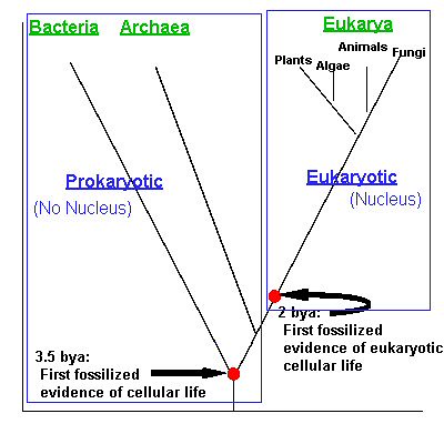
We ended the previous lecture at the beginning of our tour of the cell, and the evolution of the nucleus of the eukaryotic cell. Indeed, the cell nucleus is what defines the eukaryote domain. Prokaryotes don't have a nucleus, but rather just a chromosome in the center of the cell, a pre-nucleus (hence the label prokaryote).
We began the new lecture with a quick review of the tree of life and the split between prokaryotes and eukaryotes, and then moved on to listing some differences between the two types of cells.
The Two Basic Cell Types
Prokaryotic � � � � � � � � � � � � � � � � � � Eukaryotic
circular* � � � � �Chromosomes � � � � � �linear*
usually 1 � � � � � � � � � � � � � � � � � � � � many
in cytoplasm � � � � � � � � � � � � � � � � � �in nucleus
70s** � � � � � � � Ribosomes � � � � � � � 80s**</td>
not present � � Membrane Bound � � � �present</td>
� � � � � � � � � � � �Organelles
1 - 10 �m � � � � � �Size � � � � � � � � � �10 - 100 �m
*circular or linear: meaning the ends of the chromosome either connect or don't connect - as they are tangled up in a little ball, it is important to remember that the overall shape is not a circle or straight line
**70s and 80s are just a reference to size, with 80s being somewhat bigger than 70s
We're going to focus on Eukaryotic cells, as that domain contains the stuff we're most familiar with: plants, animals, algae, and fungi.
Eukaryotic Cell Structure
Before we get started here, let me point you to a really beautiful CGI animation of the workings of a cell. It was made by a company called XVIVO for Harvard University, and is titled The Inner Life of a Cell. It's about 8 minutes long, so watch the video, we'll discuss some of what is going on in that video, and then I'll remind you to go back and watch the video again at the end of our tour of the cell.
(The video is so amazingly well done that even the perpetrators of the Intelligent Design Creationism Hoax were so impressed with it that one of their "Leading Lights", Dr. Dr. (He's got two!!!) William A. Dembski by name, was busted having swiped it, clipped the credits, changed the title, and was using it in his "talks". Then he lied about it, and was busted in that lie too. Meanwhile, Ben Stein's utter failure of a piece of crap propaganda garbage called Expelled, got exposed for plagiarizing Inner Life as well. Such high standards of morality and integrity! Liars and thieves, the lot of them.)
(The wikipedia article has some decent diagrams of these structures, so I'm going to use them instead of my own here for illustrative purposes.)

1. Nucleus
In most eukaryotes, it is the largest structure in the cell. It's surrounded by a double membrane called the nuclear envelope. The envelope has tiny openings in it called pores.
It contains the chromosomes ---> Long molecules of DNA coiled around proteins ---> They are very, very long, but coiled up. They might be long enough to reach across a desk, but they have to fit inside the nucleus of the cell. A human being has 23 pairs of these things inside the nucleus of the cell. That big mass of 23 pairs of chromosomes is called chromatin.
Also inside the nucleus is the nucleolus ---> manufactures ribosomal RNA (rRNA). One or sometimes more than one in the nucleus.
2. Ribosomes
Sites of protein synthesis. Consists of 2 subunits of rRNA + protein We'll cover this more in Unit 4, later in the semester.
There are two groups of Ribosomes in the cell, the free and the bound (or fixed).
The free ribosomes, as the name implies, float freely in the cytoplasm, while the bound ribosomes are attached (or bound, duh) to membranes (the outer membrane of the nucleus or the outer membrane of the endoplasmic reticulum.
The other major difference is that the free ribosomes produce proteins for use in the cytoplasm of the cell, like enzymes to begin the breakdown of sugars. The bound ribosomes produce proteins for use elsewhere. Insulin is a good example of this. It is produced inside the cells of the pancreas, but used elsewhere to regulate the sugar level.
3. Endoplasmic Reticulum, or ER
endo - "inside"
plasmic - "cytoplasm"
reticulum - "net"
The ER surrounds the nucleus and is continuous with the outer membrane of the nucleus. It's divided into two parts:
Rough ER ---> has bound ribosomes, and thus looks rough. The Rough ER produces proteins for export to other organelles or other cells.
Smooth ER ---> does not have bound ribosomes, and thus looks smooth. The Smooth ER synthesizes lipids, stores Calcium (Ca2+), detoxifies chemicals ---> the enzymes inside are especially important in the liver, for example.
Proteins leave the ER via a transport vesicle. This is actually a very cool process whereby the protein presses up against the membrane of the ER, then pushes further and further out, like an erupting zit, until it forms a big blister on the outside of the ER. Eventually, the blister itself breaks free and acts like a bubble around the protein as it travels to the Golgi Apparatus.
4. Golgi Apparatus
The golgi apparatus looks kind of like a stack of pancakes or coins. When a transport vesicle touches the golgi apparatus, the membrane of the transport vesicle fuses with the membrane of the golgi apparatus. Then the blistering pimple process is repeated in reverse, and the protein is delivered inside the golgi apparatus. That's just frackin' cool.
The protein enters by transport vesicle on one end of the golgi apparatus, called the cis (or receiving) side. It will eventually leave via the trans (or shipping) side. (This is where the term cis comes from in relation to discussions with transsexual folks, if you've heard that term bandied about. Cis folks are just non-trans folks. Now you know. Next time, do your own homework.)
Inside the golgi apparatus, the protein is modified for whatever task it needs to perform. It may have sugars added or sections of the protein might be removed, etc.
The protein which entered on the cis side and was then modified in the golgi apparatus is then ready for shipping from the trans side. It's packaged up and shipped via the transport vesicle mechanism, just like before.
The transport vesicle has a protein on the outside that works like an address label, telling the vesicle where the delivery is to be made. It might go directly to the plasma membrane and exit via the same process, or it can travel to another organelle within the cell and be delivered via the same process, or it might just stay in the cytoplasm and become another membrane bound organelle, depending on its pupose.
Lysosomes are a good example of that last.
5. Lysosomes
A lysosome is a vesicle filled with hydrolytic enzymes (digestive)
Hydrolytic ---> hydrolysis (that's convenient, huh?)
The enzymes use hydrolysis to digest, or break down, stuff the cell needs broken down, like carbohydrates or proteins, etc. Different lysosomes digest different things. They are the cell's recycling guys.
They might recycle worn out organelles, breaking them down into their constituent parts to be transported and reused within the cell.
They might digest bacterial infections, etc.
Lysosomes normally digest cancerous cells.
Lysosomes can tell a cell to suicide if damaged. They also tells some cells to suicide in the normal process of development. The webs between the fingers and toes of an embryo are digested this way.
And with that, this lecture ran out of time. We picked up the discussion of eukaryotic cell structures with the next lecture, taking up mitochondria, chloroplasts, the cytoskeleton, and cilia and flagella.
--------------
“Why do creationists have such a hard time with commas?
Linky“. ~ Steve Story, Legend
|